Open
  • 4502 Riverstone Blvd, Texas 77459 , US
  • Mon - Fri 8.00 - 18.00. Saturday & Sunday CLOSED

Fully Equipped Clinic

State-Of-The-Art Facilities and Utilises

Get Fast Result

Satisfied Patients

Personalised HealthCare Service

Quality Diagnostic Treatment

Category Archives: Medical

Addressing Impingement Issues After Total Ankle Replacement

Addressing Impingement Issues After Total Ankle Replacement

As total ankle replacements (TARs) become more prevalent, it is essential for surgeons to be able to properly assess and address complications. Impingement of the bone or soft tissues can be a significant cause of pain after TAR. With this in mind, the authors provide salient diagnostic insights and offer perspective on conservative and surgical treatments for impingement, including open and arthroscopic repair. 

Total ankle replacement (TAR) is becoming an increasingly viable treatment method for ankle arthritis. Researchers have shown that TAR is a non-inferior option to ankle arthrodesis and recent data even demonstrates a trend toward better quality of life in patients who had TAR in comparison to those who had a tibiotalar arthrodesis.1-3 Due to continued improvements in technique and implant design, there has been an increase of published data on long-term survivorship of total ankle arthroplasty.4 Despite these improvements, there are still common postoperative complications surgeons may need to address.4-6

A common but often aggravating complication is gutter pain secondary to soft tissue or bony impingement. Multiple total ankle designs reportedly cause symptomatic gutter impingement in patients post-operatively.7-11 In a 2013 study involving four different ankle replacement systems and a total of 489 procedures, Schubert and colleagues reported a seven percent incidence of symptomatic gutter impingement.12 The exact etiology behind painful malleolar gutters following TAR remains unclear but the causes seem to be complex and multifactorial in nature. Potential inciting factors include technical error, ectopic bone formation, implant design, oversized components, inadequate ligamentous balancing, undercorrection of varus/valgus deformity or component loosening.9,12-15 

Pertinent Pearls In The Diagnosis Of Ankle Impingement After TAR 

The diagnosis of true ankle impingement following total ankle replacement hinges on history and physical exam in combination with radiographic evidence. The pain is localized upon palpation of the medial and lateral gutters. Additionally, eliciting pain with range of motion of the ankle joint in the sagittal and coronal planes may help localize the area of impingement. Symptoms of focal gutter pain usually present three to six months following implantation as the patient increases activity.14 

One then correlates the clinical findings with radiographic evidence, most commonly in the form of heterotopic ossification or abutment of the talar implant against the malleoli. Although osteophytes and ectopic bone ossification may be common following TAR, studies show that heterotopic ossification may not be associated with functional outcomes.16,17 Therefore, it is important to assess the clinical relevance of ossifications in relation to gutter pain. Heterotopic ossification may or may not coincide with synovial impingement following total ankle replacement.18

When there is uncertainty, utilizing other diagnostic tools may be beneficial. Employing computed tomography (CT) can allow surgeons to further assess the positioning of the prosthetic component and osseous impingements. Computed tomography is preferable to magnetic resonance imaging (MRI) because artifacts secondary to the metallic components may impede detailed assessment.19 

Diagnostic injection with local anesthetic is another useful tool in localizing the area of pain. Due to varus or valgus deformity in the ankle joint, patients often have limited use of the posterior tibial or peroneal tendons prior to ankle replacement. After correction of the deformity, patients may commonly experience inflammation and pain in those tendons because of increased activity. A diagnostic injection within the ankle joint or tendon sheaths can help delineate whether the pain is extra-articular in nature or secondary to impingement.

Conservative Treatment For Impingement After TAR: What You Need To Know 

Before considering another surgery to address gutter impingement pain following total ankle replacement, one should exhaust conservative treatments. In the early post-operative period, the patient may aggravate gutter pain from increasing activity in concurrence with post-operative inflammation. Patients may benefit from a few weeks of offloading with immobilization, rest or bracing.

Intra-articular corticosteroid injections offer another conservative treatment that can help decrease inflammation in the capsular tissues of the ankle joint. With this reduced inflammation, the reduced pressure from the thickened capsular tissue may help relieve the impingement within the medial or lateral gutters. It is important to clean the outside of the ankle prior to injection with betadine or chlorhexidine gluconate swabs so as to avoid infection to the prostheses. Also, one should avoid contacting the needle with the metallic implant as this can accelerate wear.

While it is possible that these conservative treatments may provide symptom relief, there are no current studies, to our knowledge, that assess the efficacy of these treatments for impingement symptoms following total ankle replacement.

Assessing Surgical Options For Post-TAR Ankle Impingement 

Prophylactic gutter resection is not part of the surgical technique with most available TAR systems. However, in our opinion and experience, certain prostheses such as the Scandinavian Total Ankle Replacement (STAR, Stryker) allow for gutter decompression through talar margin resection. Schuberth and colleagues found that patients who had prophylactic gutter debridement had a significantly lower incidence of secondary gutter resection than those who did not.12 

Gaudot and colleagues have also identified mobile-bearing implant systems as a potential cause of malleolar gutter pain because of the excessive subluxation of the polyethylene insert.19 Thus, adopting a fixed-bearing implant design may avoid gutter impingement. Yet the evidence is unclear on whether there is a significant difference between the two types of prostheses.

If a surgeon encounters painful malleolar gutters following TAR, it is paramount to identify the underlying cause for proper treatment. Technical error leading to malpositioning of the prosthesis is a common cause of gutter impingement pain (see top image to the left). In cases in which pain is due to malpositioned prostheses, surgeons may need to perform periprosthetic osteotomies (supramalleolar or inframalleolar) or revisional arthroplasty (see bottom image to left) to correct the deformity. The goal is to realign the joint in order to take pressure off of the symptomatic gutter. In instances of mild malposition of the prothesis, upsizing of the polyethylene insert is an option to alleviate symptoms through increased separation of the tibial and talar components.14 However, one must carefully consider the potential loss in range of motion.

Osseous overgrowth causing impingement may also attribute to aseptic loosening of the talar component.20 In such instances, a CT scan can provide a detailed assessment of the bone stock beneath the talar component. Bone cyst curettage and bone grafting may be beneficial in conjunction with resection of the osseous overgrowth. Cyst recurrence is common and complete filling of each individual cyst is often difficult.21 Therefore, revisional arthroplasty or even ankle arthrodesis may be the only solutions.

If one addresses malalignment or aseptic loosening of the prosthesis appropriately, one can perform malleolar gutter debridement through an arthroscopic or open approach.22-24 A 2.9 mm, 70-degree scope is ideal for malleolar gutter debridement. Use of an ankle distractor is the surgeon’s preference.22-24 Utilizing a combination of the shaver and grasper, the surgeon should continue to debride the gutters until there is distinct visualization of clear space between the talar bone/component and the malleoli. Once the debridement is complete, one should be able to fit the shaver within the gutters without difficulty. The advantage of arthroscopic debridement is a quicker return to activity as opposed to the open approach. However, there is significant risk of damaging the ankle components with the arthroscopic instruments due to a confined space to use the instrumentation. It is important to avoid contact to the metallic components with the shaver or burr. One must also be aware of the reflection off of the metallic component when entering the ankle joint with the shaver or burr (see top image to the right). If the surgeon is not cognizant of the orientation of the instrumentation, he or she may unintentionally damage the ankle components, and leave inflammatory debris within the ankle joint.

Debridement through an open approach allows for easier visualization and consequently a more thorough debridement of the malleolar gutters. An open approach may also decrease operating time. In instances of posterior gutter impingement secondary to ectopic ossification, an open arthrotomy is the preferred treatment option. This allows for direct visualization of the neurovascular structures that are in close proximity and difficult to avoid through an arthroscopic approach. For the open approach, one makes the incision directly over the painful gutter, taking care to protect neurovascular and tendinous structures. The surgeon can employ osteotomes and electric burrs to resect osteophytes and ectopic ossifications.

Postoperatively, patients that undergo debridement through an open ankle arthrotomy should be non-weightbearing in a posterior splint for approximately two weeks with subsequent physical therapy and progression back to regular shoes. Conversely, the patients that have arthroscopic debridement may immediately perform protected weightbearing and begin physical therapy after the first post-operative visit at one week.

Concluding Thoughts 

Total ankle prostheses continue to show improvement in long-term survivorship with each evolution of implant design. Accordingly, as prostheses show increasing longevity, complications will perpetually arise following total ankle replacement. Impingement issues after total ankle replacement are a common but complex complication that require careful consideration for management. Detailed preoperative and postoperative assessments of the patient are necessary in order to properly treat symptomatic gutter impingement. Surgeons should understand the underlying causes contributing to painful impingement so they can properly address them in conjunction with gutter debridement.

Dr. Chu is a Foot and Ankle Fellow with Coordinated Health-Lehigh Valley in Bethlehem, Pa. 

Dr. Brigido is the Director of the Coordinated Health-Lehigh Valley Fellowship in Bethlehem, Pa. He is a Fellow of the American College of Foot and Ankle Surgeons, and a Diplomate of the American Board of Podiatric Surgery. Dr. Brigido is board-certified in foot surgery and rearfoot/ankle surgery. 

References

1. Haddad SL, Coetzee JC, Estok R, Fahrbach K, Banel D, Nalysnyk L. Intermediate and long-term outcomes of total ankle arthroplasty and ankle arthrodesis. A systematic review of the literature. J Bone Joint Surg Am. 2007;89(9):1899-1905.

2. Saltzman CL, Mann RA, Ahrens JE, et al. Prospective controlled trial of STAR total ankle replacement versus ankle fusion: initial results. Foot Ankle Int. 2009;30(7):579-596.

3. Dalat F, Trouillet F, Fessy MH, Bourdin M, Besse JL. Comparison of quality of life following total ankle arthroplasty and ankle arthrodesis: retrospective study of 54 cases. Orthop Traumatol Surg Res. 2014;100(7):761-766.

4. Clough T, Bodo K, Majeed H, Davenport J, Karski M. Survivorship and long-term outcome of a consecutive series of 200 Scandinavian Total Ankle Replacement (STAR) implants. Bone Joint J. 2019;101-B(1):47-54.

5. Rippstein PF, Huber M, Naal FD. Management of specific complications related to total ankle arthroplasty. Foot Ankle Clin N Am. 2012;17:707-717.

6. Jonck JH, Myerson MS. Revision total ankle replacement. Foot Ankle Clin N Am. 2012;17(4):687-706.

7. Koivu H, Kohonen I, Mattila K, Loyttyniemi E, Tiusanen H. Long-term results of Scandinavian Total Ankle Replacement. Foot Ankle Int. 2017;38(7):723-731.

8. Cerrato R, Myerson MS. Total ankle replacement: the Agility LP prosthesis. Foot Ankle Clin. 2008;13(3):485-494.

9. Rippstein PF, Huber M, Coetzee JC, Naal FD. Total ankle replacement with use of a new three-component implant. J Bone Joint Surg Am. 2011;93(15):1426-1435.

10. Harston A, Lazarides AL, Adams SB Jr, DeOrio JK, Easley ME, Nunley JA 2nd. Midterm outcomes of a fixed-bearing total ankle arthroplasty with deformity analysis. Foot Ankle Int. 2017;38(12):1295-1300.

11. Nunley JA, Adams SB, Easley ME, DeOrio JK. Prospective randomized trial comparing mobile-bearing and fixed-bearing total ankle replacement. Foot Ankle Int. 2019;40(11):1239- 1248.

12. Schuberth JM, Babu NS, Richey JM, Christensen JC. Gutter impingement after total ankle arthroplasty. Foot Ankle Int. 2013;34(3):329- 337.

13. Gross CE, Adams SB, Easley M, Nunley JA 2nd, DeOrio JK. Surgical treatment of bony and soft-tissue impingement in total ankle arthroplasty. Foot Ankle Spec. 2017;10(1):37-42.

14. Schuberth JM, Wood DA, Christensen JC. Gutter impingement in total ankle arthroplasty. Foot Ankle Spec. 2016;9(2):145-158.

15. Krause FG, Windolf M, Bora B, Penner MJ, Wing KJ, Younger ASE. Impact of complications in total ankle replacement and ankle arthrodesis analyzed with a validated outcome measurement. J Bone Joint Surg Am. 2011;93(9):830-839.

16. Choi WJ, Lee JW. Heterotopic ossification after total ankle arthroplasty. J Bone Joint Surg Br. 2011;93(11):1508-1512.

17. Bemenderfer TB, Davis WH, Anderson RB, et al. Heterotopic ossification in total ankle arthroplasty: case series and systematic review. J Foot Ankle Surg. 2020. [Epub ahead of print] Available at: https://www.jfas.org/article/ S1067-2516(19)30452-1/fulltext . Published January 16, 2020. Accessed April 14, 2020.

18. Besse J, Bevernage BD, Leemrijse T. Revision of total ankle replacements. Tech Foot Ankle Surg. 2011;10(4):176-188.

19. Gaudot F, Colombier J-A, Bonnin M, Judet T. A controlled, comparative study of a fixed-bearing versus mobile-bearing ankle arthroplasty. Foot Ankle Int. 2014;35(2):131-140.

20. Younger A, Penner M, Wing K. Mobile-bearing total ankle arthroplasty. Foot Ankle Clin. 2008;13(3):496-508.

21. Besse J-L, Lienhart C, Fessy M-H. Outcomes following cyst curettage and bone grafting for the management of periprosthetic cystic evolution after AES total ankle replacement. Clin Podiatr Med Surg. 2013;30(2):157-170.

22. Richardson AB, DeOrio JK, Parekh SG. Arthroscopic debridement: effective treatment for impingement after total ankle arthroplasty. Curr Rev Musculoskelet Med. 2012;5(2)171-175.

23. Shirzad K, Viens NA, DeOrio JK. Arthroscopic treatment of impingement after total ankle arthroplasty: technique tip. Foot Ankle Int. 2011;32(7):727-729.

24. Kim BS, Choi WJ, Kim J, Lee JW. Residual pain due to soft-tissue impingement after uncomplicated total ankle replacement. Bone Joint J. 2013;95-B(3):378-383.

May 04, 2020
Pages: 26 – 29
By Anson K. Chu, DPM, AACFAS and Stephen A. Brigido, DPM, FACFAS

Could COVID Toes Be An Emerging Acro-Ischemia Symptom Of The COVID-19 Virus?

By Nicholas A. Campitelli, DPM, FACFAS and Kelly Kubiak DPM

COVID-19, caused by the novel coronavirus named SARS-CoV-2, causes a variety of clinical symptoms with the most common symptoms being a dry cough, fever, myalgia and fatigue.1 Less common symptoms include dyspnea, sputum production and diarrhea. However, as the COVID-19 virus continues to spread across the world, new information about the disease is emerging all the time.

One now hears the term COVID toes being noted when patients who have the COVID-19 virus present with extremity symptoms. These patients may or may not carry an official diagnosis of COVID-19. These patients may present with a digital ischemic appearance of purplish or red lesions on their toe(s) that are often painful.2 However, one could easily confuse the presentation of such symptoms for frostbite, Raynaud’s disease or chilblains. Most reports of such a phenomenon are seen primarily in younger populations with or without other symptoms.3

The exact cause of these symptoms is still unknown. One prominent theory involves a likely underrecognized vascular component to the disease.4 The COVID-19 virus is known to attack cells in the lung via the angiotensin converting enzyme 2 (ACE2) receptor. The ACE2 receptor is not limited to just the lungs. It is also found in other organs including the heart, kidney and intestines. The ACE2 receptor is also found on endothelial cells that line vessels throughout the whole circulatory system, including the very small vessels in the toes.4 Researchers out of the Pathology and Cardiology Departments from University Hospital Zurich, in Zurich, Switzerland speculate that the virus attaching in these small vessels results in the vascular symptoms now known as COVID toes.4

What Recent Case Reports Reveal About COVID Toes

As information on the novel coronavirus continues to evolve, more research on COVID toes may emerge. In a recent report out of China, Zhang and colleagues discussed seven critical patients with the COVID-19 virus, who had an average age of 59 years and clinical symptoms including finger/toe cyanosis, skin bullae and dry gangrene to the digits. These patients also reportedly had prolonged prothrombin time (PT), an elevated D-dimer level and diagnosed disseminated intravascular coagulation (DIC). Five of the seven patients ended up dying from the COVID-19 virus.

However, most reports on COVID toes come from various news media and seem to be in younger age groups with many of these patients not having any respiratory symptoms.3 A press release from the French National Union of Dermatologists and Venereologists warns of skin manifestations of COVID-19 that the group classifies as acrosyndromes.2 This group defines symptoms as the appearance of pseudo-frostbite, a sudden appearance of persistent and sometimes painful redness, and transient hive lesions on the fingers and/or toes.2

In a recent case study out of Italy from the International Federation of Podiatrists, Mazzotta and Troccoli describe self-healing lesions in children and adolescents, and believe the etiology is vascular in nature.3 Kerri Purdy, MD, FRCPC, president of the Canadian Dermatology Association, also agrees with a vascular origin.6 In a recent interview, Dr. Purdy stated that the presentation is similar to chilblains but she believes the etiology is vascular, not thermal, in nature. She attributes it to small vessel blockages as emerging evidence points to the COVID-19 virus contributing to a hypercoagulable state.6

Physicians in France and Spain also report lower extremity symptoms in various younger populations.2,3 As the aforementioned report out of China shows, COVID toe is not limited to the young but may possibly be the only symptom present in a patient with the COVID-19 virus.5

Our Experience With A Possible Presentation Of COVID-Related Pedal Symptoms

Here one can see a photograph of the patient taken at initial presentation on April 6, 2020. Her chief concern was severely painful reddish and purple lesions to her toes bilaterally. 
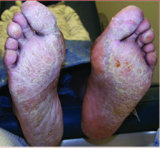
On April 6, 2020, a 13-year-old female presented to the office complaining of severely painful reddish and purple lesions to her toes bilaterally (see top two photos to right). Her symptoms began several weeks Here one can see a photograph of the patient taken at initial presentation on April 6, 2020. Her chief concern was severely painful reddish and purple lesions to her toes bilaterally. earlier and an ER physician originally treated this as cellulitis with an antibiotic. The condition eventually spread to multiple toes with blisters developing on some of the lesions (see next two photos to right). The pain was so severe the patientHere one can see a photo taken prior to presentation to the author's office, on March 31, 2020. Her symptoms were present for a few weeks, with pain making shoe gear intolerable. could not tolerate shoes.

The initial presentation was consistent with Raynaud’s disease as it was almost certainly some Here one can see a photograph taken by the patient's mother one day prior to presentation at the author's office. Even with treatment by the ER for presumed cellulitis, the lesions progressed and some even blistered.type of vasculopathy. The patient denied trauma and did not exhibit any signs or symptoms of infection. The patient had palpable dorsalis pedis and posterior tibial pulses, a sluggish capillary refill time and toes cool to the touch consistent with Raynaud’s disease. The family shared this suspicion as they noted a family history of Raynaud’s disease. At this time, this seemed to be the most likely diagnosis. We dispensed a prescription for nitroglycerin paste for the patient’s pain and symptoms.

Over ten days after presentation to the author's office, the patient related improvement in symptoms, as seen in these photos.Ten days later, the patient reported an improvement in her symptoms and clinical presentation, which was confirmed with pictures sent by the patient’s mother (see next two photos to right).

Over ten days after presentation to the author's office, the patient related improvement in symptoms, as seen in these photos.At this point in time, similar symptoms began to appear in reports of children around the world connected to COVID-19. Further questioning of the patient and her mother confirmed that the patient had a serious flu-like condition the previous month. There were also siblings in the household who had exhibited a fever, sore throat and cough approximately two weeks in duration. These siblings also tested negative for influenza and strep. When the patient began experiencing exhaustion and shortness of breath, she never had testing for influenza due to her siblings’ negative status. She did, however, test negative for mononucleosis. Her pediatrician prescribed an antibiotic and an inhaler. She did not receive a COVID-19 test.

Anecdotally, we learned through social media of a 13-year-old male from the same school of the first patient who exhibited similar symptoms and painful complaints about his toes. His symptoms had a six-week duration and consisted of erythema Here one can see a photo of a 13-year-old male from same school of the first patient who exhibited similar symptoms and painful complaints about his toes. His symptoms had a six-week duration and consisted of erythema and pain to his toes. and pain to his toes (see bottom two images to right). The erythema eventually progressed to purpuric-appearing lesions on all of the toes very similar in nature to the previous patient. His Here one can see a photo of a 13-year-old male from same school of the first patient who exhibited similar symptoms and painful complaints about his toes. His symptoms had a six-week duration and consisted of erythema and pain to his toes. pediatrician prescribed oral steroids three weeks after the initial presentation of symptoms and this treatment eventually allowed the patient to tolerate shoe gear. This patient displayed no clinical symptoms of the COVID-19 virus and had no other pertinent findings such as fever or dermatological lesions elsewhere. Accordingly, the patient was not tested for COVID-19 at that time.

In Conclusion

The aforementioned cases provide anecdotal evidence of two patients in the same geographic area who presented with symptoms that are possibly consistent with COVID toes albeit without a confirmed diagnosis of the COVID-19 virus. Both patients were in their early teens and early reports have suggested that COVID toes appear to be most prevalent in this age group.3 Both patients described color changes and a painful presentation with four to six weeks of symptoms before noting improvement. Only one of the patients exhibited crusted lesions as noted in an aforementioned report out of France.2 One patient had other symptoms suggestive of the COVID-19 virus and the other patient did not. This is consistent with similar findings in another recent report out of Spain that noted COVID toe in both symptomatic and asymptomatic patients.7 While these authors recommended topical corticosteroid treatment for patients with these lesions, other cautions exist regarding the use of systemic steroids in patients with the COVID-10 virus so practitioners should exercise caution in this population.7,8 

For the presented patients above, improvement occurred with nitroglycerin paste and topical steroids respectively.  It may be prudent to also suggest that similar patients exercise caution and self-quarantine due to the possible association with the COVID-19 virus.

At the present time, there is limited, if any, true scientific literature to guide clinical decision making in the diagnosis of these questionable COVID toes. This presents difficulty in presenting a possible diagnosis yet to be proven scientifically or backed with peer-reviewed literature. With that said, there is evidence to suggest that the two aforementioned patients who presented with pain, red-to-blue colored lesions and vasculitis to their toes could possibly have had COVID toes. The symptom timeline along with the presence of the virus in the United States supports this. Certainly, more research is necessary to specifically correlate known COVID-19 status and COVID toe presentation before we can confirm the true etiology and association of COVID toes.

Dr. Campitelli is the Director of the Podiatric Residency Program at the Western Reserve Hospital in Cuyahoga Falls, Ohio. He is an Adjunct Clinical Professor at the Kent State University School of Podiatric Medicine

Dr. Kubiak is a third-year podiatric surgery resident at the Western Reserve Hospital in Cuyahoga Falls, Ohio. 

References

  1. Huang C, Wang Y, Li X, et al. Clinical features of patients infected with 2019 novel coronavirus in Wuhan, China. Lancet. 2020;395(10223):497-506.
  2. La Revue du Praticien. Covid revealing acrosyndromes. Available at: https://drogunlana.com/2020/04/30/the-benifits-of-middle-age-fitness/ .  Accessed April 27, 2020.
  3. Mazzotta F, Troccoli T. Acute acro-ischemia in the child at the time of COVID-19. International Federation of Podiatrists. Available at: https://www.fip-ifp.org/wp-content/uploads/2020/04/acroischemia-ENG.pdf. Accessed April 27, 2020.
  4. Varga Z, Flammer AJ, Steiger P, et al. Endothelial cell infection and endotheliitis in COVID-19. Lancet. 2020. Available at: https://doi.org/10.1016/S0140-6736(20)30937-5. Accessed April 27, 2020.
  5. Zhang Y, Cao W, Xiao M, et al. Clinical and coagulation characteristics of 7 patients with critical COVID-2019 pneumonia and acro-ischemia. Chinese J Hematol. 2020;41(0):E006.
  6. Young L. ‘COVID toes’ could be another symptom of coronavirus infection: experts. Global News. Available at: https://globalnews.ca/news/6848644/covid-toes-skin-rash-coronavirus-symptom/ . Published April 21, 2020. Accessed April 27, 2020.
  7. Consejo General de Colegios Oficiales de Podólogos de España:  COVID-19 Compatible Case Register.  Available at: https://drogunlana.com/2020/04/30/the-benifits-of-middle-age-fitness/ . Accessed April 27, 2020.
  8. World Health Organization. Clinical management of severe acute respiratory infection when COVID-19 is suspected. Available at: https://www.who.int/publications-detail/clinical-management-of-severe-acute-respiratory-infection-when-novel-coronavirus-(ncov)-infection-is-suspected . Accessed April 29, 2020.
April 30, 2020
By Nicholas A Campitelli DPM FACFAS

What Role Does Shoe Cushioning Play In Running Injuries?

Injury risk increases when running in harder shoes as opposed to more cushioned shoes, according to a recent study in the American Journal of Sports Medicine.

In the study, researchers assessed 848 healthy runners over a period of six months with the runners providing data on running activity and any injuries reducing or interrupting running activity for at least seven days. The runners received one of two shoe prototypes to use with pre-determined global stiffness parameters (soft versus hard). Runners in the harder shoes had a higher injury risk. However, after stratifying for body mass, researchers found that the protection afforded by more shoe cushioning only applied to lighter runners (less than 62.8 kg for females, less than 78.2 kg for males).

Kevin Kirby, DPM relates that he believes nearly all runners benefit from some level of cushioning in their running shoe.

He recommends more firm midsoles for heavier runners or those who suffer from pronation-related injuries. Additionally, Dr. Kirby notes that dual-density midsoles that are more firm medially often assist runners with pronation-related pathology. Conversely, Dr. Kirby feels more cushioned shoes are a better fit for lighter runners with more stable feet.

“The idea is to match the shoe midsole to the runner’s weight, running style and running surface,” says Dr. Kirby, an Adjunct Associate Professor within the Department of Applied Biomechanics at the California School of Podiatric Medicine at Samuel Merritt University in Oakland, Calif. “In addition, the runner’s personal experience and injury patterns with other running shoe designs should also be taken into account.”

Alicia Canzanese, DPM also feels that shock absorption is an important feature for most runners and recommends an element of this for most distance runners. She maintains that midsole cushioning is more important for those with cavus foot, older athletes, those with a history of stress injury and runners who primarily run on pavement.

Dr. Kirby points out that for higher-mileage runners, training in more than one style or type of running shoe is beneficial.

“Two, three or four pairs of different running shoes may be helpful … so that different loading patterns will occur within the feet and lower extremities … to hopefully decrease the risk of running injury,” explains Dr. Kirby.

Dr. Kirby commends the relatively large study population and says the results make good clinical sense. He does, however, point out that those studied were very low-mileage runners, logging less than seven miles a week on average. Dr. Kirby feels this makes it difficult to extrapolate results to medium- or high-mileage runners. Additionally, he notes that the two prototype shoes are not commercially available, which challenges a meaningful comparison to running shoes on the market. Lastly, Dr. Kirby says there was a lack of clinical evaluation of the running injuries as the injuries were self-reported in the study.

Dr. Canzanese states the results of this study support the importance of cushioning to protect runners from repetitive load stress. However, there are many specifications not addressed in this study that help determine what shoe is appropriate for a runner, says Dr. Canzanese, a member of the Pennsylvania Podiatric Medical Association Executive Board.


Study Evaluates Long-Term Survival Rate Of Youngswick Osteotomies

By Jennifer Spector, DPM, FACFAS, Associate Editor

Will patients undergoing a Youngswick osteotomy for moderate hallux rigidus eventually need a first MPJ fusion?

In a recently published retrospective study in the Journal of Foot and Ankle Surgery, researchers evaluated the use of a Youngswick osteotomy in 61 patients with stage II and III hallux rigidus. Over a mean follow-up time of 54.8 months, all patients showed improved Foot and Ankle Outcome Scores (FAOS) with final postoperative scores greater than 75 points.

Nearly half of the study patients demonstrated radiographic worsening of the first MPJ over the follow-up period but no patient progressed to arthrodesis. The study authors concluded that the Youngswick osteotomy provides satisfactory long-term functional, pain and patient satisfaction outcomes, even in some patients up to 13 years postoperatively.

Jeffrey S. Boberg, DPM, FACFAS has used the Youngswick osteotomy for over two decades, primarily in earlier stages of hallux limitus/rigidus with a long first metatarsal or in later stages, when the proximal phalanx sits plantar to the first metatarsal head.

“I have never had to revise a Youngswick correction,” shares Dr. Boberg, who is in private practice in O’Fallon, Missouri. “I do not claim this is universally successful … but I did a several-year follow-up on (my patients with the procedure) in the late 1990s and none of the patients regretted having the surgery.”

Dr. Boberg feels the benefits of the Youngswick procedure are the joint decompression and alteration of the first MPJ mechanics.

“In hallux rigidus, the sesamoids are frozen. Instead of the phalanx gliding over the metatarsal head, the sesamoids become a pivot point for the phalangeal base,” points out Dr. Boberg, a faculty member of the Podiatry Institute. “This results in the dorsal aspect of the base of the proximal phalanx impacting the dorsal half of the first metatarsal head (resulting in cartilage loss and pain in this same area). By plantarflexing and shortening the metatarsal, there is some small increase in sesamoid motion but significantly less contact between the phalangeal base and the first metatarsal head.”

Dr. Boberg notes that other than those in the earliest stages of hallux limitus/rigidus, most patients do not see any appreciable increase in joint motion after a Youngswick osteotomy.

“The goal of the procedure should be pain reduction, not increased range of motion,” maintains Dr. Boberg. “(My) patients function without pain, demonstrate propulsion and limited metatarsalgia, but with a stiff joint. They walk as if they had a fusion but with a more rapid recovery and less morbidity than they would have had with an arthrodesis.”


How Should One Initially Treat Minimally Displaced Lisfranc Injuries?

By Jennifer Spector, DPM, FACFAS, Associate Editor

What are the consequences of treating minimally displaced Lisfranc injuries conservatively?

Over five years, researchers assessed 26 patients that sustained minimally displaced Lisfranc injuries and had a subsequent non-surgical treatment course. The collected data included radiological outcomes and patient-reported outcome scores at least one-year post-injury, according to the study published recently in Foot and Ankle International. 

Study authors found that 54 percent of the patients sustained further injury displacement with a median time to displacement of 18 days. At a mean follow-up time of 54 months, researchers noted that the patient-reported outcomes were comparable between the group that remained minimally displaced and the group that underwent surgical intervention to address the additional displacement despite the delay in surgical attention.

Jacob Wynes, DPM, FACFAS, relates that an understanding of the literature is quite important to best manage these types of injuries. Regardless of one’s preferred surgical approach, Dr. Wynes notes that one cannot underestimate the high rate of tarsometatarsal osteoarthritis when minimally displaced Lisfranc injuries are left untreated.

“In my opinion, … diastasis should prompt the foot and ankle specialist to not only stress the lateral tarsometatarsal joint but also assess first tarsometatarsal joint instability,” notes Dr. Wynes, an Assistant Professor of Orthopaedics at the University of Maryland School of Medicine.

In his practice, Dr. Wynes considers bridge plating of the first tarsometatarsal joint and screw fixation from the medial cuneiform to the second metatarsal base (a “home run screw”) when there is diastasis between the first and second metatarsals along with first tarsometatarsal joint instability. He also employs another screw from the lateral base of the third metatarsal with an oblique orientation toward the intermediate cuneiform.

In regard to patients treated non-operatively for minimally displaced Lisfranc injuries, Dr. Wynes relates he has yet to see a patient respond favorably. While he often sees patients referred for additional opinions in his practice, he shares that it is difficult to obtain patient agreement for surgical intervention after initial conservative treatment. Dr. Wynes points out that the longer there is diastasis, the longer the patient experiences uneven joint contact.

“The authors (of this study) still recommend surgery if secondary instability results,” notes Dr. Wynes. “I would argue that latent instability and likely more advanced joint disease after displacement (then warrants) primary arthrodesis as opposed to ORIF. This could allow for improved functional and patient-reported outcomes.”

Hi, How Can We Help You?