Open
  • 4502 Riverstone Blvd, Texas 77459 , US
  • Mon - Fri 8.00 - 18.00. Saturday & Sunday CLOSED

Fully Equipped Clinic

State-Of-The-Art Facilities and Utilises

Get Fast Result

Satisfied Patients

Personalised HealthCare Service

Quality Diagnostic Treatment

Author Archives: drogunlana

Addressing Impingement Issues After Total Ankle Replacement

Addressing Impingement Issues After Total Ankle Replacement

As total ankle replacements (TARs) become more prevalent, it is essential for surgeons to be able to properly assess and address complications. Impingement of the bone or soft tissues can be a significant cause of pain after TAR. With this in mind, the authors provide salient diagnostic insights and offer perspective on conservative and surgical treatments for impingement, including open and arthroscopic repair. 

Total ankle replacement (TAR) is becoming an increasingly viable treatment method for ankle arthritis. Researchers have shown that TAR is a non-inferior option to ankle arthrodesis and recent data even demonstrates a trend toward better quality of life in patients who had TAR in comparison to those who had a tibiotalar arthrodesis.1-3 Due to continued improvements in technique and implant design, there has been an increase of published data on long-term survivorship of total ankle arthroplasty.4 Despite these improvements, there are still common postoperative complications surgeons may need to address.4-6

A common but often aggravating complication is gutter pain secondary to soft tissue or bony impingement. Multiple total ankle designs reportedly cause symptomatic gutter impingement in patients post-operatively.7-11 In a 2013 study involving four different ankle replacement systems and a total of 489 procedures, Schubert and colleagues reported a seven percent incidence of symptomatic gutter impingement.12 The exact etiology behind painful malleolar gutters following TAR remains unclear but the causes seem to be complex and multifactorial in nature. Potential inciting factors include technical error, ectopic bone formation, implant design, oversized components, inadequate ligamentous balancing, undercorrection of varus/valgus deformity or component loosening.9,12-15 

Pertinent Pearls In The Diagnosis Of Ankle Impingement After TAR 

The diagnosis of true ankle impingement following total ankle replacement hinges on history and physical exam in combination with radiographic evidence. The pain is localized upon palpation of the medial and lateral gutters. Additionally, eliciting pain with range of motion of the ankle joint in the sagittal and coronal planes may help localize the area of impingement. Symptoms of focal gutter pain usually present three to six months following implantation as the patient increases activity.14 

One then correlates the clinical findings with radiographic evidence, most commonly in the form of heterotopic ossification or abutment of the talar implant against the malleoli. Although osteophytes and ectopic bone ossification may be common following TAR, studies show that heterotopic ossification may not be associated with functional outcomes.16,17 Therefore, it is important to assess the clinical relevance of ossifications in relation to gutter pain. Heterotopic ossification may or may not coincide with synovial impingement following total ankle replacement.18

When there is uncertainty, utilizing other diagnostic tools may be beneficial. Employing computed tomography (CT) can allow surgeons to further assess the positioning of the prosthetic component and osseous impingements. Computed tomography is preferable to magnetic resonance imaging (MRI) because artifacts secondary to the metallic components may impede detailed assessment.19 

Diagnostic injection with local anesthetic is another useful tool in localizing the area of pain. Due to varus or valgus deformity in the ankle joint, patients often have limited use of the posterior tibial or peroneal tendons prior to ankle replacement. After correction of the deformity, patients may commonly experience inflammation and pain in those tendons because of increased activity. A diagnostic injection within the ankle joint or tendon sheaths can help delineate whether the pain is extra-articular in nature or secondary to impingement.

Conservative Treatment For Impingement After TAR: What You Need To Know 

Before considering another surgery to address gutter impingement pain following total ankle replacement, one should exhaust conservative treatments. In the early post-operative period, the patient may aggravate gutter pain from increasing activity in concurrence with post-operative inflammation. Patients may benefit from a few weeks of offloading with immobilization, rest or bracing.

Intra-articular corticosteroid injections offer another conservative treatment that can help decrease inflammation in the capsular tissues of the ankle joint. With this reduced inflammation, the reduced pressure from the thickened capsular tissue may help relieve the impingement within the medial or lateral gutters. It is important to clean the outside of the ankle prior to injection with betadine or chlorhexidine gluconate swabs so as to avoid infection to the prostheses. Also, one should avoid contacting the needle with the metallic implant as this can accelerate wear.

While it is possible that these conservative treatments may provide symptom relief, there are no current studies, to our knowledge, that assess the efficacy of these treatments for impingement symptoms following total ankle replacement.

Assessing Surgical Options For Post-TAR Ankle Impingement 

Prophylactic gutter resection is not part of the surgical technique with most available TAR systems. However, in our opinion and experience, certain prostheses such as the Scandinavian Total Ankle Replacement (STAR, Stryker) allow for gutter decompression through talar margin resection. Schuberth and colleagues found that patients who had prophylactic gutter debridement had a significantly lower incidence of secondary gutter resection than those who did not.12 

Gaudot and colleagues have also identified mobile-bearing implant systems as a potential cause of malleolar gutter pain because of the excessive subluxation of the polyethylene insert.19 Thus, adopting a fixed-bearing implant design may avoid gutter impingement. Yet the evidence is unclear on whether there is a significant difference between the two types of prostheses.

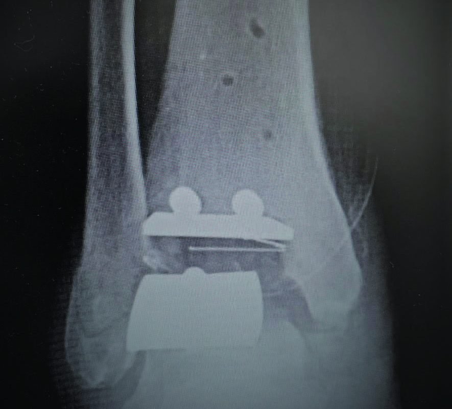
If a surgeon encounters painful malleolar gutters following TAR, it is paramount to identify the underlying cause for proper treatment. Technical error leading to malpositioning of the prosthesis is a common cause of gutter impingement pain (see top image to the left). In cases in which pain is due to malpositioned prostheses, surgeons may need to perform periprosthetic osteotomies (supramalleolar or inframalleolar) or revisional arthroplasty (see bottom image to left) to correct the deformity. The goal is to realign the joint in order to take pressure off of the symptomatic gutter. In instances of mild malposition of the prothesis, upsizing of the polyethylene insert is an option to alleviate symptoms through increased separation of the tibial and talar components.14 However, one must carefully consider the potential loss in range of motion.

Osseous overgrowth causing impingement may also attribute to aseptic loosening of the talar component.20 In such instances, a CT scan can provide a detailed assessment of the bone stock beneath the talar component. Bone cyst curettage and bone grafting may be beneficial in conjunction with resection of the osseous overgrowth. Cyst recurrence is common and complete filling of each individual cyst is often difficult.21 Therefore, revisional arthroplasty or even ankle arthrodesis may be the only solutions.

If one addresses malalignment or aseptic loosening of the prosthesis appropriately, one can perform malleolar gutter debridement through an arthroscopic or open approach.22-24 A 2.9 mm, 70-degree scope is ideal for malleolar gutter debridement. Use of an ankle distractor is the surgeon’s preference.22-24 Utilizing a combination of the shaver and grasper, the surgeon should continue to debride the gutters until there is distinct visualization of clear space between the talar bone/component and the malleoli. Once the debridement is complete, one should be able to fit the shaver within the gutters without difficulty. The advantage of arthroscopic debridement is a quicker return to activity as opposed to the open approach. However, there is significant risk of damaging the ankle components with the arthroscopic instruments due to a confined space to use the instrumentation. It is important to avoid contact to the metallic components with the shaver or burr. One must also be aware of the reflection off of the metallic component when entering the ankle joint with the shaver or burr (see top image to the right). If the surgeon is not cognizant of the orientation of the instrumentation, he or she may unintentionally damage the ankle components, and leave inflammatory debris within the ankle joint.

Debridement through an open approach allows for easier visualization and consequently a more thorough debridement of the malleolar gutters. An open approach may also decrease operating time. In instances of posterior gutter impingement secondary to ectopic ossification, an open arthrotomy is the preferred treatment option. This allows for direct visualization of the neurovascular structures that are in close proximity and difficult to avoid through an arthroscopic approach. For the open approach, one makes the incision directly over the painful gutter, taking care to protect neurovascular and tendinous structures. The surgeon can employ osteotomes and electric burrs to resect osteophytes and ectopic ossifications.

Postoperatively, patients that undergo debridement through an open ankle arthrotomy should be non-weightbearing in a posterior splint for approximately two weeks with subsequent physical therapy and progression back to regular shoes. Conversely, the patients that have arthroscopic debridement may immediately perform protected weightbearing and begin physical therapy after the first post-operative visit at one week.

Concluding Thoughts 

Total ankle prostheses continue to show improvement in long-term survivorship with each evolution of implant design. Accordingly, as prostheses show increasing longevity, complications will perpetually arise following total ankle replacement. Impingement issues after total ankle replacement are a common but complex complication that require careful consideration for management. Detailed preoperative and postoperative assessments of the patient are necessary in order to properly treat symptomatic gutter impingement. Surgeons should understand the underlying causes contributing to painful impingement so they can properly address them in conjunction with gutter debridement.

Dr. Chu is a Foot and Ankle Fellow with Coordinated Health-Lehigh Valley in Bethlehem, Pa. 

Dr. Brigido is the Director of the Coordinated Health-Lehigh Valley Fellowship in Bethlehem, Pa. He is a Fellow of the American College of Foot and Ankle Surgeons, and a Diplomate of the American Board of Podiatric Surgery. Dr. Brigido is board-certified in foot surgery and rearfoot/ankle surgery. 

References

1. Haddad SL, Coetzee JC, Estok R, Fahrbach K, Banel D, Nalysnyk L. Intermediate and long-term outcomes of total ankle arthroplasty and ankle arthrodesis. A systematic review of the literature. J Bone Joint Surg Am. 2007;89(9):1899-1905.

2. Saltzman CL, Mann RA, Ahrens JE, et al. Prospective controlled trial of STAR total ankle replacement versus ankle fusion: initial results. Foot Ankle Int. 2009;30(7):579-596.

3. Dalat F, Trouillet F, Fessy MH, Bourdin M, Besse JL. Comparison of quality of life following total ankle arthroplasty and ankle arthrodesis: retrospective study of 54 cases. Orthop Traumatol Surg Res. 2014;100(7):761-766.

4. Clough T, Bodo K, Majeed H, Davenport J, Karski M. Survivorship and long-term outcome of a consecutive series of 200 Scandinavian Total Ankle Replacement (STAR) implants. Bone Joint J. 2019;101-B(1):47-54.

5. Rippstein PF, Huber M, Naal FD. Management of specific complications related to total ankle arthroplasty. Foot Ankle Clin N Am. 2012;17:707-717.

6. Jonck JH, Myerson MS. Revision total ankle replacement. Foot Ankle Clin N Am. 2012;17(4):687-706.

7. Koivu H, Kohonen I, Mattila K, Loyttyniemi E, Tiusanen H. Long-term results of Scandinavian Total Ankle Replacement. Foot Ankle Int. 2017;38(7):723-731.

8. Cerrato R, Myerson MS. Total ankle replacement: the Agility LP prosthesis. Foot Ankle Clin. 2008;13(3):485-494.

9. Rippstein PF, Huber M, Coetzee JC, Naal FD. Total ankle replacement with use of a new three-component implant. J Bone Joint Surg Am. 2011;93(15):1426-1435.

10. Harston A, Lazarides AL, Adams SB Jr, DeOrio JK, Easley ME, Nunley JA 2nd. Midterm outcomes of a fixed-bearing total ankle arthroplasty with deformity analysis. Foot Ankle Int. 2017;38(12):1295-1300.

11. Nunley JA, Adams SB, Easley ME, DeOrio JK. Prospective randomized trial comparing mobile-bearing and fixed-bearing total ankle replacement. Foot Ankle Int. 2019;40(11):1239- 1248.

12. Schuberth JM, Babu NS, Richey JM, Christensen JC. Gutter impingement after total ankle arthroplasty. Foot Ankle Int. 2013;34(3):329- 337.

13. Gross CE, Adams SB, Easley M, Nunley JA 2nd, DeOrio JK. Surgical treatment of bony and soft-tissue impingement in total ankle arthroplasty. Foot Ankle Spec. 2017;10(1):37-42.

14. Schuberth JM, Wood DA, Christensen JC. Gutter impingement in total ankle arthroplasty. Foot Ankle Spec. 2016;9(2):145-158.

15. Krause FG, Windolf M, Bora B, Penner MJ, Wing KJ, Younger ASE. Impact of complications in total ankle replacement and ankle arthrodesis analyzed with a validated outcome measurement. J Bone Joint Surg Am. 2011;93(9):830-839.

16. Choi WJ, Lee JW. Heterotopic ossification after total ankle arthroplasty. J Bone Joint Surg Br. 2011;93(11):1508-1512.

17. Bemenderfer TB, Davis WH, Anderson RB, et al. Heterotopic ossification in total ankle arthroplasty: case series and systematic review. J Foot Ankle Surg. 2020. [Epub ahead of print] Available at: https://www.jfas.org/article/ S1067-2516(19)30452-1/fulltext . Published January 16, 2020. Accessed April 14, 2020.

18. Besse J, Bevernage BD, Leemrijse T. Revision of total ankle replacements. Tech Foot Ankle Surg. 2011;10(4):176-188.

19. Gaudot F, Colombier J-A, Bonnin M, Judet T. A controlled, comparative study of a fixed-bearing versus mobile-bearing ankle arthroplasty. Foot Ankle Int. 2014;35(2):131-140.

20. Younger A, Penner M, Wing K. Mobile-bearing total ankle arthroplasty. Foot Ankle Clin. 2008;13(3):496-508.

21. Besse J-L, Lienhart C, Fessy M-H. Outcomes following cyst curettage and bone grafting for the management of periprosthetic cystic evolution after AES total ankle replacement. Clin Podiatr Med Surg. 2013;30(2):157-170.

22. Richardson AB, DeOrio JK, Parekh SG. Arthroscopic debridement: effective treatment for impingement after total ankle arthroplasty. Curr Rev Musculoskelet Med. 2012;5(2)171-175.

23. Shirzad K, Viens NA, DeOrio JK. Arthroscopic treatment of impingement after total ankle arthroplasty: technique tip. Foot Ankle Int. 2011;32(7):727-729.

24. Kim BS, Choi WJ, Kim J, Lee JW. Residual pain due to soft-tissue impingement after uncomplicated total ankle replacement. Bone Joint J. 2013;95-B(3):378-383.

May 04, 2020
Pages: 26 – 29
By Anson K. Chu, DPM, AACFAS and Stephen A. Brigido, DPM, FACFAS

Could COVID Toes Be An Emerging Acro-Ischemia Symptom Of The COVID-19 Virus?

By Nicholas A. Campitelli, DPM, FACFAS and Kelly Kubiak DPM

COVID-19, caused by the novel coronavirus named SARS-CoV-2, causes a variety of clinical symptoms with the most common symptoms being a dry cough, fever, myalgia and fatigue.1 Less common symptoms include dyspnea, sputum production and diarrhea. However, as the COVID-19 virus continues to spread across the world, new information about the disease is emerging all the time.

One now hears the term COVID toes being noted when patients who have the COVID-19 virus present with extremity symptoms. These patients may or may not carry an official diagnosis of COVID-19. These patients may present with a digital ischemic appearance of purplish or red lesions on their toe(s) that are often painful.2 However, one could easily confuse the presentation of such symptoms for frostbite, Raynaud’s disease or chilblains. Most reports of such a phenomenon are seen primarily in younger populations with or without other symptoms.3

The exact cause of these symptoms is still unknown. One prominent theory involves a likely underrecognized vascular component to the disease.4 The COVID-19 virus is known to attack cells in the lung via the angiotensin converting enzyme 2 (ACE2) receptor. The ACE2 receptor is not limited to just the lungs. It is also found in other organs including the heart, kidney and intestines. The ACE2 receptor is also found on endothelial cells that line vessels throughout the whole circulatory system, including the very small vessels in the toes.4 Researchers out of the Pathology and Cardiology Departments from University Hospital Zurich, in Zurich, Switzerland speculate that the virus attaching in these small vessels results in the vascular symptoms now known as COVID toes.4

What Recent Case Reports Reveal About COVID Toes

As information on the novel coronavirus continues to evolve, more research on COVID toes may emerge. In a recent report out of China, Zhang and colleagues discussed seven critical patients with the COVID-19 virus, who had an average age of 59 years and clinical symptoms including finger/toe cyanosis, skin bullae and dry gangrene to the digits. These patients also reportedly had prolonged prothrombin time (PT), an elevated D-dimer level and diagnosed disseminated intravascular coagulation (DIC). Five of the seven patients ended up dying from the COVID-19 virus.

However, most reports on COVID toes come from various news media and seem to be in younger age groups with many of these patients not having any respiratory symptoms.3 A press release from the French National Union of Dermatologists and Venereologists warns of skin manifestations of COVID-19 that the group classifies as acrosyndromes.2 This group defines symptoms as the appearance of pseudo-frostbite, a sudden appearance of persistent and sometimes painful redness, and transient hive lesions on the fingers and/or toes.2

In a recent case study out of Italy from the International Federation of Podiatrists, Mazzotta and Troccoli describe self-healing lesions in children and adolescents, and believe the etiology is vascular in nature.3 Kerri Purdy, MD, FRCPC, president of the Canadian Dermatology Association, also agrees with a vascular origin.6 In a recent interview, Dr. Purdy stated that the presentation is similar to chilblains but she believes the etiology is vascular, not thermal, in nature. She attributes it to small vessel blockages as emerging evidence points to the COVID-19 virus contributing to a hypercoagulable state.6

Physicians in France and Spain also report lower extremity symptoms in various younger populations.2,3 As the aforementioned report out of China shows, COVID toe is not limited to the young but may possibly be the only symptom present in a patient with the COVID-19 virus.5

Our Experience With A Possible Presentation Of COVID-Related Pedal Symptoms

Here one can see a photograph of the patient taken at initial presentation on April 6, 2020. Her chief concern was severely painful reddish and purple lesions to her toes bilaterally. On April 6, 2020, a 13-year-old female presented to the office complaining of severely painful reddish and purple lesions to her toes bilaterally (see top two photos to right). Her symptoms began several weeks Here one can see a photograph of the patient taken at initial presentation on April 6, 2020. Her chief concern was severely painful reddish and purple lesions to her toes bilaterally. earlier and an ER physician originally treated this as cellulitis with an antibiotic. The condition eventually spread to multiple toes with blisters developing on some of the lesions (see next two photos to right). The pain was so severe the patientHere one can see a photo taken prior to presentation to the author's office, on March 31, 2020. Her symptoms were present for a few weeks, with pain making shoe gear intolerable. could not tolerate shoes.

The initial presentation was consistent with Raynaud’s disease as it was almost certainly some Here one can see a photograph taken by the patient's mother one day prior to presentation at the author's office. Even with treatment by the ER for presumed cellulitis, the lesions progressed and some even blistered.type of vasculopathy. The patient denied trauma and did not exhibit any signs or symptoms of infection. The patient had palpable dorsalis pedis and posterior tibial pulses, a sluggish capillary refill time and toes cool to the touch consistent with Raynaud’s disease. The family shared this suspicion as they noted a family history of Raynaud’s disease. At this time, this seemed to be the most likely diagnosis. We dispensed a prescription for nitroglycerin paste for the patient’s pain and symptoms.

Over ten days after presentation to the author's office, the patient related improvement in symptoms, as seen in these photos.Ten days later, the patient reported an improvement in her symptoms and clinical presentation, which was confirmed with pictures sent by the patient’s mother (see next two photos to right).

Over ten days after presentation to the author's office, the patient related improvement in symptoms, as seen in these photos.At this point in time, similar symptoms began to appear in reports of children around the world connected to COVID-19. Further questioning of the patient and her mother confirmed that the patient had a serious flu-like condition the previous month. There were also siblings in the household who had exhibited a fever, sore throat and cough approximately two weeks in duration. These siblings also tested negative for influenza and strep. When the patient began experiencing exhaustion and shortness of breath, she never had testing for influenza due to her siblings’ negative status. She did, however, test negative for mononucleosis. Her pediatrician prescribed an antibiotic and an inhaler. She did not receive a COVID-19 test.

Anecdotally, we learned through social media of a 13-year-old male from the same school of the first patient who exhibited similar symptoms and painful complaints about his toes. His symptoms had a six-week duration and consisted of erythema Here one can see a photo of a 13-year-old male from same school of the first patient who exhibited similar symptoms and painful complaints about his toes. His symptoms had a six-week duration and consisted of erythema and pain to his toes. and pain to his toes (see bottom two images to right). The erythema eventually progressed to purpuric-appearing lesions on all of the toes very similar in nature to the previous patient. His Here one can see a photo of a 13-year-old male from same school of the first patient who exhibited similar symptoms and painful complaints about his toes. His symptoms had a six-week duration and consisted of erythema and pain to his toes. pediatrician prescribed oral steroids three weeks after the initial presentation of symptoms and this treatment eventually allowed the patient to tolerate shoe gear. This patient displayed no clinical symptoms of the COVID-19 virus and had no other pertinent findings such as fever or dermatological lesions elsewhere. Accordingly, the patient was not tested for COVID-19 at that time.

In Conclusion

The aforementioned cases provide anecdotal evidence of two patients in the same geographic area who presented with symptoms that are possibly consistent with COVID toes albeit without a confirmed diagnosis of the COVID-19 virus. Both patients were in their early teens and early reports have suggested that COVID toes appear to be most prevalent in this age group.3 Both patients described color changes and a painful presentation with four to six weeks of symptoms before noting improvement. Only one of the patients exhibited crusted lesions as noted in an aforementioned report out of France.2 One patient had other symptoms suggestive of the COVID-19 virus and the other patient did not. This is consistent with similar findings in another recent report out of Spain that noted COVID toe in both symptomatic and asymptomatic patients.7 While these authors recommended topical corticosteroid treatment for patients with these lesions, other cautions exist regarding the use of systemic steroids in patients with the COVID-10 virus so practitioners should exercise caution in this population.7,8 

For the presented patients above, improvement occurred with nitroglycerin paste and topical steroids respectively.  It may be prudent to also suggest that similar patients exercise caution and self-quarantine due to the possible association with the COVID-19 virus.

At the present time, there is limited, if any, true scientific literature to guide clinical decision making in the diagnosis of these questionable COVID toes. This presents difficulty in presenting a possible diagnosis yet to be proven scientifically or backed with peer-reviewed literature. With that said, there is evidence to suggest that the two aforementioned patients who presented with pain, red-to-blue colored lesions and vasculitis to their toes could possibly have had COVID toes. The symptom timeline along with the presence of the virus in the United States supports this. Certainly, more research is necessary to specifically correlate known COVID-19 status and COVID toe presentation before we can confirm the true etiology and association of COVID toes.

Dr. Campitelli is the Director of the Podiatric Residency Program at the Western Reserve Hospital in Cuyahoga Falls, Ohio. He is an Adjunct Clinical Professor at the Kent State University School of Podiatric Medicine

Dr. Kubiak is a third-year podiatric surgery resident at the Western Reserve Hospital in Cuyahoga Falls, Ohio. 

References

  1. Huang C, Wang Y, Li X, et al. Clinical features of patients infected with 2019 novel coronavirus in Wuhan, China. Lancet. 2020;395(10223):497-506.
  2. La Revue du Praticien. Covid revealing acrosyndromes. Available at: https://drogunlana.com/2020/04/30/the-benifits-of-middle-age-fitness/ .  Accessed April 27, 2020.
  3. Mazzotta F, Troccoli T. Acute acro-ischemia in the child at the time of COVID-19. International Federation of Podiatrists. Available at: https://www.fip-ifp.org/wp-content/uploads/2020/04/acroischemia-ENG.pdf. Accessed April 27, 2020.
  4. Varga Z, Flammer AJ, Steiger P, et al. Endothelial cell infection and endotheliitis in COVID-19. Lancet. 2020. Available at: https://doi.org/10.1016/S0140-6736(20)30937-5. Accessed April 27, 2020.
  5. Zhang Y, Cao W, Xiao M, et al. Clinical and coagulation characteristics of 7 patients with critical COVID-2019 pneumonia and acro-ischemia. Chinese J Hematol. 2020;41(0):E006.
  6. Young L. ‘COVID toes’ could be another symptom of coronavirus infection: experts. Global News. Available at: https://globalnews.ca/news/6848644/covid-toes-skin-rash-coronavirus-symptom/ . Published April 21, 2020. Accessed April 27, 2020.
  7. Consejo General de Colegios Oficiales de Podólogos de España:  COVID-19 Compatible Case Register.  Available at: https://drogunlana.com/2020/04/30/the-benifits-of-middle-age-fitness/ . Accessed April 27, 2020.
  8. World Health Organization. Clinical management of severe acute respiratory infection when COVID-19 is suspected. Available at: https://www.who.int/publications-detail/clinical-management-of-severe-acute-respiratory-infection-when-novel-coronavirus-(ncov)-infection-is-suspected . Accessed April 29, 2020.
April 30, 2020
By Nicholas A Campitelli DPM FACFAS
Diabetic Foot Ulcers

Can Remote Ischemic Conditioning Promote Healing In Diabetic Foot Ulcers?

The prevalence of diabetes is on the rise worldwide. As podiatrists and diabetic foot experts, we know the deleterious effects of diabetes on the tissues of the lower extremity. Among diabetes-related complications, the treatment and management of diabetic foot ulcers (DFUs) remains major challenges for patients, caregivers and health-care systems alike. Multiple disrupted physiologic processes, including decreases in cellular signaling and growth factor responsiveness, lead to microvascular dysfunction and diminished peripheral blood flow that can contribute to the lack of healing in people with DFUs.

Successful translation of novel therapeutic modalities into clinical algorithms for DFU management may fulfill an unmet need that is of increasing importance given the global diabetes epidemic. There is an abundance of clinical evidence that remote ischemic conditioning is cardioprotective but can it provide the same protection to the microvascular circulation of patients with diabetes, and accordingly help increase healing rates in patients with DFUs?

Researchers initially studied remote ischemic conditioning (RIC) as a potential protective strategy for cardiac function. In 1986, Murry and colleagues discovered that short repetitive bouts of occlusion and reperfusion of a coronary artery in dogs subsequently protected the heart against a myocardial infarction.1 In 1993, Przyklenk and team conducted a study that is considered the first evidence for the remote application of tissue conditioning.2 This study showed that brief controlled periods of occlusion and reperfusion of a canine coronary artery also protected remote cardiac tissue not directly supplied by this artery when subjected to a subsequent sustained ischemic episode.

Drawing upon on this data, researchers began investigating whether remote ischemic conditioning provided analogous benefits to patients with tissue ischemia injuries. Subsequent clinical studies in human models have concluded that remote ischemic conditioning is safe, well-tolerated and produces a systemic phenomenon that has beneficial effects in other organs such as lung, liver, kidney, intestines and the brain as well as skeletal muscle tissues.3,4


What The Research Reveals About Endothelial Dysfunction, Microvascular Disease And Remote Ischemic Conditioning

Ischemic Conditioning Promote Healing In Diabetic Foot Ulcers?While the prevailing thinking is that the etiology of diabetic microvascular disease is multifactorial, a consistent finding in patients with diabetes is endothelial dysfunction.5 There is a known correlation between the long-term effects of elevated glucose levels and the alteration of endothelial cell function.5 An impairment in the formation of vasodilators such as nitric oxide along with increases in the formation of several vasoconstrictors speed the progression of microvascular disease.6 It is generally accepted that hyperglycemia resulting from uncontrolled diabetes leads to an impairment of nitric oxide production and activity.6,7 Prolonged elevated glucose levels generate oxidants in smooth muscle that may diminish nitric oxide signaling, decreasing the responsiveness of endothelium-dependent vasodilation, especially in the microcirculation.6,7 The effects of this cascade of events are decreased functional perfusion and tissue hypoxia in the lower extremity, particularly the feet, complicating DFU healing.

There is scientific evidence indicating that one effect of remote ischemic conditioning is an increase in nitric oxide production.7 One hypothesis is that the repetitive inflation and deflation of a blood pressure cuff has a shearing effect on the vasculature that results in the release of nitric oxide.7 Researchers have suggested that remote ischemic conditioning may contribute to improved endothelial function, resulting in enhanced vascular performance.7 Reversal of tissue hypoxia and increases in peripheral circulation could potentially improve wound healing, especially in patients who are not candidates for other vascular interventions.

In a 2011 study, Kraemer and colleagues treated 27 healthy patients with remote ischemic conditioning and examined tissue oxygenation and capillary blood flow in the anterolateral aspect of the left thigh.8 After patients had three five-minute cycles of remote ischemic conditioning to the contralateral upper arm, researchers found statistically significant increases from baseline measurements of 29 and 35 percent in tissue oxygenation and capillary blood flow respectively. These increases occurred during the third reperfusion phase.8 The results of this study appear to support evidence of increased microvascular blood flow in the lower extremity, furthering the idea that remote ischemic conditioning could potentially aid in DFU healing.

In a 2014 double-blind, prospective, randomized study involving 40 patients with aseptic and infected DFUs, Shaked and colleagues assessed the efficacy of remote ischemic conditioning as an adjunct to standard of care treatment.9 Applying blood pressure cuffs to both arms of all the patients, researchers inflated and deflated the cuffs for three five-minute cycles. The study group had their cuffs inflated to 200 mmHg while the control group had their cuffs inflated to 10 mmHg. The patients in the study group had remote ischemic conditioning treatments every two weeks and were followed for a total of six weeks. For the patients who completed the study, nine out of 22 patients (41 percent) in the treatment arm achieved complete wound healing in comparison to zero out of 12 patients in the control group.


Could Remote Ischemic Conditioning Have An Impact In Limb Salvage Protocols? 

Most clinical remote ischemic conditioning treatment studies use a standard blood pressure cuff or similar device applied to the upper or lower extremity to produce the cycles of non-lethal ischemia. Treatment typically consists of three or four cycles that medical personnel can administer over approximately 40 minutes by inflating and deflating the blood pressure cuff every five minutes. Clinicians reportedly achieve the greatest effects with treatments every 72 hours or two to three times a week.10

The occlusion pressure needs to be at least 25 mm above the patient’s systolic pressure, which averages 125 mmHg but can be much higher.10 Therefore, medical personnel need to determine the patient’s systolic pressure first and monitor it throughout treatment. One option is to go arbitrarily high on all patients but even a set pressure of 200 mm leaves nine percent of patients with DFUs uncovered and is very uncomfortable for all patients, potentially reducing compliance.10 Thus, integrating such treatments into regular clinical practice would be costly in time and medical staff resources as well as patient satisfaction.Remote-Ischemic-Conditioning

An emerging modality, the HomeCuff Wound Therapy device (LifeCuff Technologies), is reportedly showing promise in early studies.11 According to the company, this automated remote ischemic conditioning device is specifically designed for home use with easy application to the arm by the patient or a caregiver. The modality operates through a single push-button, which is pre-programmed to deliver an automated 40-minute treatment cycle without the need for medical personnel, thus reducing the cost of treatment. Unlike standard blood pressure cuffs that can only apply a single set pressure, the HomeCuff Wound Therapy device applies variable occlusive pressure based on intermittent readings of extremity blood pressure from software within the cuff.10,11 This facilitates the delivery of remote ischemic conditioning at the most effective yet comfortable level.10,11

The device has a built-in electronic monitoring system that collects and transmits adherence to treatment regimen data and vital sign values to a secure and HIPAA-compliant database. Early case studies showed promising results with the use of the HomeCuff Wound Therapy device two to three times weekly to treat patients with DFUs.10


How Remote Ischemic Conditioning Helped Heal An Ulcer Of Three Months In Duration 

A 68-year-old male presented with a three-month history of a neuropathic ulcer (4.75 cm2) to the left first metatarsal head (see first photo above). His past medical history included non-insulin-dependent diabetes mellitus (NIDDM), diabetic neuropathy, stage 3 cardiovascular disease, cirrhosis, anemia and hepatic encephalopathy. The patient previously tried and failed multiple advanced wound care therapies before using the HomeCuff Wound Therapy device. He began 40-minute treatments with this modality three times weekly. Secondary wound dressings consisted of Drawtex® hydroconductive wound dressing (Beier Drawtex Healthcare), ABD padding, rolled gauze and Coban. The wound completely healed in seven weeks (see second photo above).


Concluding Thoughts 

Preliminary case study results utilizing remote ischemic conditioning as an adjunctive therapy in the treatment of hard-to-heal DFUs appear promising. Further research into remote ischemic conditioning is necessary in order to prove its utility in wound care. A full understanding of preclinical data as well as the methods and mechanisms involved with remote ischemic conditioning will help wound care clinicians determine when and if to employ remote ischemic conditioning as an adjunctive therapy. Randomized clinical trials may help facilitate the translation of such new technologies to a clinically feasible paradigm for home use.

Dr. Cole is the Medical Director of the Wound Care Center at University Hospitals Ahuja Medical Center in Beachwood, Ohio. She is also an Adjunct Professor and Director of Wound Care Research at the Kent State University School of Podiatric Medicine. 

Ms. Coe is a Clinical Research Coordinator in Wound Care Research at the Kent State University College of Podiatric Medicine. Since 2015, she has been a Certified Clinical Research Professional through the Society of Clinical Research Associates (SOCRA). 

References

1. Murry CE, Jennings RB, Reimer KA. Preconditioning with ischemia: a delay of lethal cell injury in ischemic myocardium. Circulation. 1986;74(5):1124-36.

2. Przyklenk K, Bauer B, Ovize M, Kloner RA, Whittaker P. Regional ischemic ‘preconditioning’ protects remote virgin myocardium from subsequent sustained coronary occlusion. Circulation. 1993;87(3):893–899.

3. Vasdekis SN, Athanasiadis D, Lazaris A, et al. The role of remote ischemic preconditioning in the treatment of atherosclerotic diseases. Brain Behav. 2013;3(6):606–616.

4. Lim SY, Hausenloy DJ. Remote ischemic conditioning: from bench to bedside. Front Physiol. 2012;3:27.

5. Avogaro A, Albiero M, Menegazzo L, de Kreutzenberg S, Fadini GP. Endothelial dysfunction in diabetes: the role of reparatory mechanisms. Diabetes Care. 2011;34 Suppl 2(Suppl 2):S285– S290.

6. Cohen RA. Role of nitric oxide in diabetic complications. Am J Ther. 2005;12(6):499–502.

7. Kimura M, Ueda K, Goto C, et al. Repetition of ischemic preconditioning augments endothelium-dependent vasodilation in humans: role of endothelium-derived nitric oxide and endothelial progenitor cells. Arterioscler Thromb Vasc Biol. 2007;27(6):1403–1410.

8. Kraemer R, Lorenzen J, Kabbani M, et al. Acute effects of remote ischemic preconditioning on cutaneous microcirculation–a controlled prospective cohort study. BMC Surg. 2011;11:32.

9. Shaked G, Czeiger D, Abu Arar A, Katz T, Harman-Boehm I, Sebbag G. Intermittent cycles of remote ischemic preconditioning augment diabetic foot ulcer healing. Wound Repair Regen. 2015;23(2):191–196.

10. Personal communication with Thomas Moore, BA, Chairman and CEO of LifeCuff Technologies, on January 9, January 11 and February 27, 2020.

11. Garratt KN, Leschinsky B. Remote ischemic conditioning: the commercial market: LifeCuff perspective. J Cardiovasc Pharmacol Ther. 2017;22(5):408–413.

April 28, 2020
Pages: 12 – 16
By Windy Cole, DPM and Stacey Coe, BA, CCRP

Keys To Diagnosing And Treating Xerotic Skin Conditions

Given the common nature of xerotic skin disorders as well as the varied array of etiologies and treatments, these authors offer a thorough review of the literature on conditions ranging from ichthyosis and atopic dermatitis to venous stasis dermatitis and asteatotic dermatitis. 

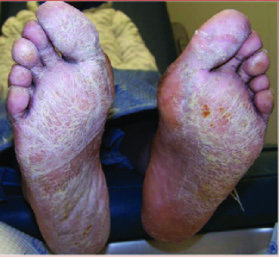
Xerosis is a very common skin disorder characterized by excessively dry skin. Other terms for this disorder include xerosis cutis and xeroderma. Xerosis can be a primary pathology associated with loss of the normal water content of the epidermis. Xerotic skin can also occur secondary to associated skin disorders and systemic disease. Underlying all xerotic skin disorders is excess water loss from the epidermis.

Skin requires a water content of 10 to 15 percent to remain intact and maintain normal function.1 Three main deficiencies in the skin lead to the development of xerosis including deficiency in natural moisturizing factors; deficiency in the skin lipids or ceramides; and deficiency in moisture in the epidermis that is mediated by aquaporin water channels.2-7 Natural moisturizing factors are isolated to the stratum corneum in high concentration in the corneocytes. These factors consist of amino acids and their derivatives including lactate, urea and inorganic salts.2 Lipids in the stratum corneum modulate water loss. Deficiencies of these cutaneous lipids can increase epidermal water loss up to 75 times that of normal skin.8

Ceramides are the main lipids in the stratum corneum. Numerous risk factors contribute to loss of cutaneous lipids and predispose individuals to develop xerotic skin disorders. This may include decreased sebaceous and sweat gland activity associated with aging; anti-androgen therapy, which decreases sebum production; exposure to degreasing agents including soaps and solvents; and exposure to dry environments.

Xerosis has variable presentation depending on its severity. Mild xerosis can exhibit accentuation of skin lines and resemble the appearance of cracked porcelain due to epidermal water loss. Xerosis affects the normal desquamation process of the epidermis, leading to the development of thin flakes on the skin surface. With more severe xerosis, one will see pruritic, dry, cracked and fissured skin. Severe xerosis can produce an inflammatory dermatitis with localized erythema and edema. Clinicians may note xerotic skin on numerous areas of the body including the lower extremity, upper extremity, abdomen and face.

Patients of increased age are at significantly higher risk of developing xerotic skin disorders.9 Sebaceous gland activity decreases significantly after 70 years of age in women and 80 years of age in men.10 Sweat gland function also declines with age.11 Skin thickness decreases with age, leading to increased water loss from the skin to the environment.12 Environmental factors are also significant risk factors for the development of xerosis. In winter months when humidity decreases, xerosis occurs much more frequently. Xerotic skin disorders are more common in dry climates with low humidity.

Basic treatment for all xerotic skin disorders aims to minimize cutaneous water loss. Lazar and Lazar identified the following methods to prevent water loss and lubricate the skin:

• reduce the frequency of bathing, showering and skin cleansing;

• increase room humidity;

• limit exposure to soaps, detergents, solvents and water;

• avoid friction from washcloths, clothing and other abrasives; and

• use emollients frequently.13

Moisturizers are a mainstay in the treatment of xerotic skin. The skin contains natural moisturizers including ceramides, glycerol, urea and lactic acid. Many moisturizers contain these elements aiming to supplement these natural moisturizing agents. Skin care products that both improve skin hydration and improve barrier function are wise choices. Specific products should contain both rehydrating and lipid-restoring components. Urea has the largest body of evidence for the treatment of xerosis.14 Combining urea with moisturizing agents and ceramides can improve its effectiveness.

Aiming to address multiple key deficiencies in skin hydration, Weber and colleagues formulated a topical formulation containing glyceryl glucoside, natural moisturizing factors and ceramide, and found it to be an effective treatment modality for xerosis.15

Addressing Asteatotic Dermatitis And Ichthyosis In The Podiatric Patient 

Asteatotic dermatitis is an inflammatory dermatitis secondary to severely xerotic skin. Other terms for this disorder include xerotic dermatitis, xerotic eczema and eczema craquelé. Asteatotic dermatitis most commonly occurs in elderly people with underlying xerosis.

Asteatotic dermatitis can be generalized or localized. Generalized disease is often associated with underlying systemic disease. Localized forms most commonly occur on the pretibial areas. Patients with asteatotic dermatitis exhibit dry, cracked and polygonal fissured skin with scaling and pruritis. Secondary erythema, edema and excoriations can develop from scratching. Fissures with superficial bleeding can occur when the skin develops cracks deep enough to damage dermal capillaries.

Known as “winter itch,” asteatotic dermatitis most commonly occurs in the winter months when environmental humidity is the lowest. Asteatotic dermatitis is prevalent in the elderly due to decreased sebaceous and sweat gland activity associated with aging. Aside from climate and age, certain medications, including diuretics, retinoids and protein kinase inhibitors, can also contribute to the development of asteatotic dermatitis.16

In addition to the preventative skin care recommended by Lazar and Lazar, topical steroid ointments under occlusion and Unna boots are treatment options for asteatotic eczema.13,17 Topical calcineurin inhibitors, including pimecrolimus and tacrolimus cream, show efficacy in the treatment of asteatotic dermatitis.18 Recently, endogenous phospholipids, N-palmitoylethanolamine and N-acetylethanolamine, that are part of the endocannabinoid system have proven to be effective treatments for asteatotic dermatitis with efficacy superior to traditional emollients.19

Ichthyosis is a group of skin disorders characterized by excessive dry, scaling skin. The name for this disorder comes from the Greek word, ichthys, meaning fish, since this disorder is known for its xerotic scales. Both inherited and acquired forms of ichthyosis exist with the most common form being ichthyosis vulgaris, an inherited autosomal-dominant disorder that commonly begins in childhood.20 Patients with ichthyosis vulgaris have xerotic skin with fine white scales. Scaling is most common on the extensor surfaces of the extremities. Acquired ichthyosis typically occurs in adults and is associated with medications that inhibit sterol synthesis in epidermal cells (nicotinic acid) or underlying systemic diseases including Hodgkin’s lymphoma, leukemia, sarcoidosis, human immunodeficiency virus (HIV), hypothyroidism, hepatitis, malabsorption and bone marrow transplantation.21 Acquired ichthyosis appears as small white scales on the extremities.

Clinicians may treat ichthyosis with topical creams and emollients to hydrate the skin and keratolytics to remove scales.

Creams containing a high percentage of urea or lactic acid can be very effective treatment options for ichthyosis.22 Oral retinoids such as acitretin (Soriatane) and isotretinoin have a general anti-keratinizing effect, and the literature suggests effectiveness in the treatment of more severe cases of ichthyosis.20

What Are The Best Approaches For Atopic And Venous Stasis Dermatitis? 

Atopic dermatitis is an inflammatory skin disorder, which is often associated with xerotic skin. This disorder presents as dry, itchy, red, swollen and cracked skin. There is often serous drainage and the presentation can vary with age. A total body distribution is more typical in infancy. For children, it is more common to see atopic dermatitis in the back of the knees and the front of the elbows. The feet and hands are the most common sites in adults.

Frequently, atopic dermatitis is associated with allergies and asthma. Several factors are thought to contribute to the development of atopic dermatitis including genetics, immune system dysfunction, environmental triggers and disruption of skin permeability. Dry skin secondary to dry climate, frequent washing and harsh chemicals increases the risk of developing atopic dermatitis.23

Treatment of atopic dermatitis varies based on the severity of the disease. Basic treatment involves avoiding aggravating environments and keeping the skin moist with moisturizers and emollients.24 Mild to moderate disease may respond to topical corticosteroids.25 Oral corticosteroids and calcineurin inhibitors are applicable for the treatment of more severe and resistant cases.23,26-28

Venous stasis dermatitis is a common inflammatory disorder affecting the skin of the lower extremities. It is frequently one of the first manifestations of chronic venous insufficiency, when retrograde blood flow through incompetent valves leads to venous hypertension and the eventual extravasation of red blood cells and ferric iron into dermal tissues. Dermal tissue changes results both directly from venous hypertension and from an inflammatory process mediated by metalloproteinases that are upregulated by ferric iron in extravasated red blood cells.29

Stasis dermatitis appears as erythematous, scaling, eczematous patches on the lower extremity. The medial ankle is the most common site, owing to its relatively poor blood supply. Skin lesions can vary in distribution from small patches to areas encompassing the entire lower leg below the knee and involving the dorsal foot. Long-standing skin lesions can present with lichenification and hyperpigmentation. Additionally, chronic venous insufficiency and hypertension can lead to skin induration and progression to lipodermatosclerosis.30

The treatment of stasis dermatitis involves management of the underlying venous insufficiency and edema. One typically treats this condition through compression therapy.29 Xerotic skin in areas of quiescent dermatitis often responds to emollients and moisturizers. Mid-potency topical steroids are applicable for short durations in the management of acute inflammation and pruritus. Long-term and high-potency topical corticosteroids are not desirable as they can lead to steroid-induced cutaneous atrophy, which can increase the risk of developing venous skin ulcerations.31,32

While topical calcineurin inhibitors are only approved for the treatment of atopic dermatitis, they are reportedly effective treatment modalities for many inflammatory skin disorders including stasis dermatitis.33,34 Tacrolimus has specifically proven effective in the treatment of stasis dermatitis.35 Maroo and colleagues found a combination of topical tacrolimus and oral doxycycline to be effective for stasis dermatitis.36

What Is The Relationship Between Systemic Disease And Xerotic Skin? 

Several systemic diseases can cause xerosis and the workup of xerotic skin changes should include consideration of underlying systemic disease. Disorders including diabetes mellitus, thyroid disease and severe renal disease are frequently associated with xerotic skin. Treatment of xerosis secondary to systemic disease typically involves management of the underlying disease state as well as symptomatic management.

It is common to observe xerotic skin in patients with diabetes mellitus. Dry skin has the potential to fissure, increasing the risk of foot ulceration and infection in patients with diabetes mellitus.37,38

The nervous system plays an important role in maintaining adequate skin hydration. Diabetic polyneuropathy affects small sympathetic nerves, resulting in atrophy of sweat glands and decreased sudomotor response.39-42

Additionally, microcirculatory disease in patients with diabetes can lead to dry, rough, atrophic skin. Namgoong and team specifically examined the effect of peripheral neuropathy and microangiopathy on skin hydration in the feet of patients with diabetes mellitus.43 These researchers found a significant correlation between skin hydration and microvascularity, but no significant correlation between skin hydration and peripheral nerve function.

Hypothyroidism is a disorder of the endocrine system in which the thyroid gland fails to produce adequate amounts of thyroid hormone. Thyroid dysfunction is more common in women and people over the age of 60. This underproduction of thyroid hormones decreases the activity of the sweat glands, resulting in dry, xerotic skin.44 Skin changes in hypothyroidism include coarse, thin, scaly skin.45 The prevailing theory is that reduction of thyroid hormone alters sterol synthesis in epidermal keratinocytes, leading to xerotic skin changes.46 Treatment of hypothyroid-associated skin changes involves treatment of the underlying endocrine disorder with thyroid hormone supplementation.

Skin disorders are also extremely common in patients with chronic renal failure (CRF) and end-stage renal disease (ESRD).47 Xerosis is the most common skin disorder associated with renal disease, reportedly occurring in over 80 percent of patients with chronic renal failure.48 When it comes to the development of xerosis in chronic renal failure and ESRD, researchers have proposed several etiologies including decreased sweat production, decreased sebum production, reduced lipids in the skin surface, altered vitamin A metabolism, loss of or reduction in epidermal water content, and disruption of the integrity of the stratum corneum.49,50

In chronic renal failure and ESRD, reduced glomerular filtration rate leads to accumulation of waste products, including urea, creatinine, sodium, calcium, and phosphate, that are some of the main agents associated with the pathogenesis of skin disease in severe renal disease.51 Patients with severe xerosis secondary to renal disease can develop ichthyosis. Moisturizers with 5-10% urea cream or 2-3% salicylic acid are options for the treatment of uremic xerosis.49,52

In Conclusion 

Xerotic skin disorders are very common and have numerous etiologies including local and systemic disease. Both age and environmental factors play significant roles in the development of these disorders. Management of xerotic skin varies based on severity and pathology, and frequently involves management of environmental risk factors, emollients and moisturizers, and treatment of underlying disease states.

Dr. Hoffman is an Attending Physician in the Department of Orthopedics at Denver Health Medical Center. She is an Assistant Professor in the Department of Orthopedics at the University of Colorado School of Medicine. She is an Attending Physician for the Highland/Presbyterian St. Luke’s Medical Center Residency Program. 

Dr. Jerabek is an Attending Physician in the Department of Orthopedics at Denver Health Medical Center. She is an Assistant Professor in the Department of Orthopedics at the University of Colorado School of Medicine. 

References

1. Pons-Guiraud A. Dry skin in dermatology: a complex physiopathology. J Eur Acad Dermatol Venereol. 2007;21 Suppl 2:1-4.

2. Rawlings AV, Scott IR, Harding CR, Bowser PA. Stratum corneum moisturization at the molecular level. J Invest Dermatol. 1994;103(5):731- 741.

3. Rawlings AV, Harding CR. Moisturization and skin barrier function. Dermatol Ther. 2004;17 Suppl 1:43-48.

4. Jungersted JM, Hellgren LI, Jemec GB, Agner T. Lipids and skin barrier function–a clinical perspective. Contact Dermatitis. 2008;58(5):255- 262.

5. Elias PM, Feingold KR. Lipids and the epidermal water barrier: metabolism, regulation, and pathophysiology. Semin Dermatol. 1992;11(2):176-182.

6. Draelos ZD. New channels for old cosmeceuticals: aquaporin modulation. J Cosmet Dermatol. 2008;7(2):83.

7. Bonte F. Skin moisturization mechanisms: new data. Ann Pharm Fr. 2011;69(3):135-141.

8. Akimoto K, Yoshikawa N, Higaki Y, Kawashima M, Imokawa G. Quantitative analysis of stratum corneum lipids in xerosis and asteatotic eczema. J Dermatol. 1993;20(1):1-6.

9. White-Chu EF, Reddy M. Dry skin in the elderly: complexities of a common problem. Clin Dermatol. 2011;29(1):37-42.

10. Pochi PE, Strauss JS, Downing DT. Age-related changes in sebaceous gland activity. J Invest Dermatol. 1979;73(1):108-111.

11. Anderson RK, Kenney WL. Effect of age on heat-activated sweat gland density and flow during exercise in dry heat. J Appl Physiol. 1987;63(3):1089-1094.

12. Farage MA, Miller KW, Elsner P, Maibach HI. Characteristics of the aging skin. Adv Wound Care (New Rochelle). 2013;2(1):5-10.

13. Lazar AP, Lazar P. Dry skin, water, and lubrication. Dermatol Clin. 1991;9(1):45-51.

14. Augustin M, Wilsmann-Theis D, Korber A, et al. Diagnosis and treatment of xerosis cutis – a position paper. J Dtsch Dermatol Ges. 2019;17 Suppl 7:3-33.

15. Weber TM, Kausch M, Rippke F, Schoelermann AM, Filbry AW. Treatment of xerosis with a topical formulation containing glyceryl glucoside, natural moisturizing factors, and ceramide. J Clinical Aesth Dermatol. 2012;5(8):29- 39.

16. Norman R. Xerosis and pruritus in the elderly—recognition and management. In: Norman R, ed. Diagnosis of Aging Skin Diseases. London: Springer London; 2008.

17. Ward S. Eczema and dry skin in older people: identification and management. Br J Community Nurs. 2005;10(10):453-456.

18. Schulz P, Bunselmeyer B, Brautigam M, Luger TA. Pimecrolimus cream 1% is effective in asteatotic eczema: results of a randomized, double-blind, vehicle-controlled study in 40 patients. J Eur Acad Dermatol Venereol. 2007;21(1):90-94.

19. Yuan C, Wang XM, Guichard A, et al. N-palmitoylethanolamine and N-acetylethanolamine are effective in asteatotic eczema: results of a randomized, double-blind, controlled study in 60 patients. Clin Interv Aging. 2014;9:1163- 1169.

20. Vahlquist A, Fischer J, Torma H. Inherited nonsyndromic ichthyoses: an update on pathophysiology, diagnosis and treatment. Am J Clin Dermatol. 2018;19(1):51-66.

21. DiGiovanna JJ, Robinson-Bostom L. Ichthyosis: etiology, diagnosis, and management. Am J Clin Dermatol. 2003;4(2):81-95.

22. Blair C. The action of a urea-lactic acid ointment in ichthyosis with particular reference to the thickness of the horny layer. Br J Dermatol. 1976;94(2):145-153.

23. Tollefson MM, Bruckner AL, Section On Dermatology. Atopic dermatitis: skin-directed management. Pediatrics. 2014;134(6):e1735-1744.

24. Varothai S, Nitayavardhana S, Kulthanan K. Moisturizers for patients with atopic dermatitis. Asian Pac J Allergy Immunol. 2013;31(2):91-98.

25. Berke R, Singh A, Guralnick M. Atopic dermatitis: an overview. Am Fam Phys. 2012;86(1):35- 42.

26. Ashcroft DM, Chen LC, Garside R, Stein K, Williams HC. Topical pimecrolimus for eczema. Cochrane Database Syst Rev. 2007(4):CD005500.

27. Cury Martins J, Martins C, Aoki V, Gois AF, Ishii HA, da Silva EM. Topical tacrolimus for atopic dermatitis. Cochrane Database Syst Rev. 2015(7):CD009864.

28. Carr WW. Topical calcineurin inhibitors for atopic dermatitis: review and treatment recommendations. Paediatr Drugs. 2013;15(4):303-310.

29. Sundaresan S, Migden MR, Silapunt S. Stasis dermatitis: pathophysiology, evaluation, and management. Am J Clin Dermatol. 2017;18(3):383-390.

30. Kirsner RS, Pardes JB, Eaglstein WH, Falanga V. The clinical spectrum of lipodermatosclerosis. J Am Acad Dermatol. 1993;28(4):623-627.

31. Wilkinson SM, English JS. Hydrocortisone sensitivity: clinical features of fifty-nine cases. J Am Acad Dermatol. 1992;27(5 Pt 1):683-687.

32. Lubach D, Bensmann A, Bornemann U. Steroid-induced dermal atrophy. Investigations on discontinuous application. Dermatologica. 1989;179(2):67-72.

33. Lin AN. Innovative use of topical calcineurin inhibitors. Dermatol Clin. 2010;28(3):535-545.

34. Wollina U. The role of topical calcineurin inhibitors for skin diseases other than atopic dermatitis. Am J Clin Dermatol. 2007;8(3):157-173.

35. Dissemond J, Knab J, Lehnen M, Franckson T, Goos M. Successful treatment of stasis dermatitis with topical tacrolimus. Vasa. 2004;33(4):260-262.

36. Maroo N, Choudhury S, Sen S, Chatterjee S. Oral doxycycline with topical tacrolimus for treatment of stasis dermatitis due to chronic venous insufficiency: A pilot study. Indian J Pharmacol. 2012;44(1):111-113.

37. Papanas N, Maltezos E. The diabetic foot: established and emerging treatments. Acta Clin Belg. 2007;62(4):230-238.

38. Boulton AJ. The diabetic foot: grand overview, epidemiology and pathogenesis. Diabetes Metab Res Rev. 2008;24 Suppl 1:S3-6.

39. Low VA, Sandroni P, Fealey RD, Low PA. Detection of small-fiber neuropathy by sudomotor testing. Muscle Nerve. 2006;34(1):57-61.

40. Tesfaye S, Boulton AJ, Dyck PJ, et al. Diabetic neuropathies: update on definitions, diagnostic criteria, estimation of severity, and treatments. Diabetes Care. 2010;33(10):2285-2293.

41. Papanas N, Ziegler D. New diagnostic tests for diabetic distal symmetric polyneuropathy. J Diabetes Complications. 2011;25(1):44-51.

42. Papanas N, Boulton AJ, Malik RA, et al. A simple new non-invasive sweat indicator test for the diagnosis of diabetic neuropathy. Diab Med. 2013;30(5):525-534.

43. Namgoong S, Yang JP, Han SK, Lee YN, Dhong ES. Influence of peripheral neuropathy and microangiopathy on skin hydration in the feet of patients with diabetes mellitus. Wounds. 2019;31(7):173-178.

44. Safer JD. Thyroid hormone action on skin. Dermatoendocrinol. 2011;3(3):211-215.

45. Kohn LT, Corrigan JM, Donaldson MS (eds). To Err is Human: Building a Safer Health System. Washington, DC: Institute of Medicine: National Academies Press; 2000. DOI: 10.17226/9728

46. Rosenberg RM, Isseroff RR, Ziboh VA, Huntley AC. Abnormal lipogenesis in thyroid hormone-deficient epidermis. J Invest Dermatol. 1986;86(3):244-248.

47. Amatya B, Agrawal S, Dhali T, Sharma S, Pandey SS. Pattern of skin and nail changes in chronic renal failure in Nepal: a hospital-based study. J Dermatol. 2008;35(3):140-145.

48. Sheikh M ML, Jahangir M. Cutaneous manifestations of chronic renal failure. J Pakistan Assn Dermatol. 2014;24(2):150-155.

49. Szepietowski JC, Reich A, Schwartz RA. Uraemic xerosis. Nephrol Dial Transplant. 2004;19(11):2709-2712.

50. Lupi O, Rezende L, Zangrando M, et al. Cutaneous manifestations in end-stage renal disease. An Bras Dermatol. 2011;86(2):319-326.

51. Galperin TA, Cronin AJ, Leslie KS. Cutaneous manifestations of ESRD. Clin J Am Soc Nephrol. 2014;9(1):201-218.

52. Kuypers DR. Skin problems in chronic kidney disease. Nat Clin Pract Nephrol. 2009;5(3):157- 170.

April 03, 2020
Pages: 36 – 41
By Kristine Hoffman DPM, FACFAS and Morgan Jerabek, DPM
Is Total Ankle Replacement Appropriate In Cases Of Severe Coronal Deformity?

Is Total Ankle Replacement Appropriate In Cases Of Severe Coronal Deformity?

Although preoperative coronal deformity greater than 20 degrees has historically been a contraindication for total ankle replacement (TAR), a recent study in the Journal of Bone and Joint Surgery contends that this is not necessarily true.

The study authors assessed 148 ankles after TAR and noted that 41 ankles had severe coronal deformity over 20 degrees. Employing radiographic and clinical evaluation, the authors compared outcomes between severe and moderate deformity groups. After a mean of 74 months follow-up, there was no significant difference in pain scores, disability scores, range of motion or complication rates between those with severe and moderate deformities. Postoperative tibiotalar angle and talar tilt angle were greater in the severe deformity group.

Ryan McMillen, DPM, FACFAS relates that he tries to perform TAR on congruent ankle joints, pointing out that implant survivorship is not the only consideration.

“It’s also about edge loading and how it develops in joints with at least 10 degrees of coronal deformity,” explains Dr. McMillen, a member of the faculty for the Western Pennsylvania Hospital Foot and Ankle Residency Program in Pittsburgh. “This can lead to a need for poly exchange or abnormal wear on the implant.”

Mark Prissel, DPM, FACFAS, shares that large valgus deformities are more challenging and may require a staged approach, especially if they are associated with a flatfoot deformity and/or deltoid insufficiency.

Dr. McMillen agrees that ligamentous balancing may be required with these larger deformities and notes that he has seen no increase in complications in the short and intermediate terms with this balancing.

While this study showed similar results among the cohorts studied, Dr. Prissel says this may not be true in lower volume centers.

“Complex TAR with large angular deformity should be performed by experienced TAR surgeons at centers of high volume,” maintains Dr. Prissel, who is in private practice with multiple locations in Ohio.

Dr. McMillen adds that a comparison to patients with low to no coronal deformity would have been interesting to see with this study. He acknowledges that this study could cause him to more closely consider a patient for TAR who has more than 10 degrees of deformity and is otherwise a strong candidate for the procedure.

Study Says Younger And Less Active Patients More Prone To Sever’s Disease

A recent study in the Journal of the American Podiatric Medical Association found that younger (mean age 9.8 years) and less active (sports sessions less than 60 minutes) patients are more likely to suffer from calcaneal apophysitis.

The study included 430 children (328 male, 102 female) aged six to 14 years old. Most of the children participated in sports a mean of 2.8 times per week with each session being 60 to 120 minutes for most respondents. In addition to the primary findings regarding age and activity level, the study authors did not identify any significant differences with regard to sex, foot posture, BMI, terrain type or type of sport.

Stephen Kominsky, DPM, FACFAS says a keen understanding of the biomechanics of Sever’s disease is key to successful outcomes. Maggie Fournier, DPM, FACFAS echoes the importance of biomechanics.

When asked about the typical patient profile in their practices with calcaneal apophysitis, both doctors have had similar clinical experiences seeing males around 11 years old in high-impact or running sports. However, age, gender and activity level may vary.

Dr. Kominsky shares that this study will not change his current patient treatment protocols.

“I believe that treatment should be broken down into activity modification, mechanical support … and non-steroidal anti-inflammatory drugs (NSAIDs),” notes Dr. Kominsky, the former Director of Podiatric Medical Education at the Washington Hospital Center in Washington, DC. “Then, based on availability, things like physical therapy, laser and stretching/yoga can be of additional benefit.”

Relating that this study reinforces her current protocol for Sever’s disease, Dr. Fournier explains that a thorough history and sound physical examination should lead to a correct diagnosis without the need for additional imaging (unless there is concern of additional or different diagnoses).

“I do not hesitate to utilize ancillary services such as those provided by athletic trainers or physical therapists,” notes Dr. Fournier, the Immediate Past President of the American Academy of Podiatric Sports Medicine. “(Calcaneal apophysitis) can be a lingering and frustrating issue due to varying responses to treatment and continued sports demands on the patient.  However, we should not hesitate to modify our treatment plans to provide the most effective care.”

Study Looks At Umbilical Tissue In DFUs With Osteomyelitis

Could cryopreserved umbilical cord be an emerging option for complex, non-healing DFUs with osteomyelitis? A recent study in Wound Repair and Regeneration evaluated the use of such tissue (TTAX01) for these complex cases.

Over a 16-week trial involving 32 patients with DFUs and underlying osteomyelitis, researchers performed initial surgical debridement and then the patients had a combination of systemic antibiotics with application of TTAX01. Patients received repeat applications of TTAX01 at no less than four-week intervals. The authors reported no major amputations and noted a 91 percent mean wound area reduction from baseline.

Eric Leonheart, DPM relates treating countless DFUs and osteomyelitis over 25 years in practice. Although he has not used umbilical cord biologics, Dr. Leonheart shares he would only use biologic graft materials in complex wounds (exposed tendon, muscle, joint and bone) that are osteomyelitis-free.

Stephanie Wu, DPM, MSc, FACFAS, a co-author of the study, says most advanced biologics are not indicated for complex, deep wounds with osteomyelitis.

“It is rare to see a trial that focuses on complicated, deep, diabetic foot ulcers that extend to muscle, capsule or bone with radiographic evidence of osteomyelitis. There is truly a need for research (such as this) to assess the efficacy of novel biologic treatments to improve and accelerate healing in these complex wounds,” says Dr. Wu, the Associate Dean of Research, a Professor of Surgery at the Dr. William M. Scholl College of Podiatric Medicine and a Professor of Stem Cell and Regenerative Medicine at the School of Graduate Medical Sciences at the Rosalind Franklin University of Medicine and Science.

Dr. Leonheart finds the study’s suggestion that TTAX01 may be a possibility for DFUs with osteomyelitis concerning, citing a lack of detailed information on infection staging, debridement and management along with a lack of control group.

“I am a firm believer in the principles of umbilical and placental biologics when it comes to augmenting compromised wound healing. However, I would not change my way of treating these complex infections based on the findings in this publication,” states Dr. Leonheart, who is affiliated with the Department of Orthopedics at Madigan Army Medical Center in Tacoma.

Dr. Wu states that it is important to note that this study is not a large-scale, randomized, controlled trial, and that one purpose of this study was to examine the operational aspects and ease of compliance with the study protocol before initiating a larger, phase 3 study.

“We look forward to the confirmation of these findings in larger studies involving randomized comparison to other treatment strategies,” adds Dr. Wu.

November 25, 2019
Pages:10 – 11
By Jennifer Spector, DPM, Associate Editor
Hi, How Can We Help You?